Main Menu
- Home
- Product Finder
- Calibration Systems
- Calibration Services
- Digital Sensing
- Industrial Vibration Calibration
- Modal and Vibration Testing
- Non-Destructive Testing
- Sound & Vibration Rental Program
- Learn
- About Us
- Contact Us
 Professor Elijah Van Houten, PhD, of the Université de Sherbrooke's Department of Mechanical Engineering in Québec, Canada, has been conducting research on alternative testing for breast cancer tumors. The current diagnosis method is a mammogram, a low-energy x-ray of breast tissue that is often reported as painful by those who receive it.
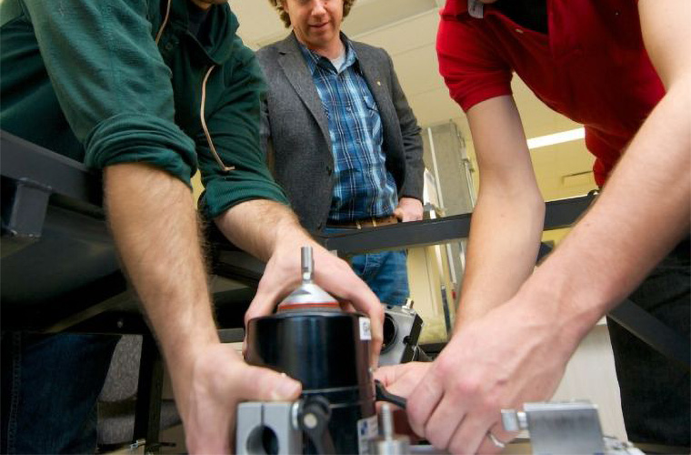
Professor Van Houten's research is using a 2025E modal shaker from The Modal Shop to perform two relatively new techniques -- elastography and elastotomography. Essentially, these techniques interpret data associated with the elasticity of soft tissue and show the contrast revealed by the presence of more rigid tissue, like tumors.
"The goal of elastography, an approach featured in Professor Van Houten's PhD research, is to make a picture of mechanical properties, especially the elasticity of soft tissue. It is known that in the case of breast cancer, women themselves detect a cancerous tumor because of its rigidity. It has been observed that tumors can be 100% to 1000% 'harder' than healthy tissue," he says.
To create the picture of breast tissue mechanical properties, Professor Van Houten uses magnetic resonance elastography. Data obtained by magnetic resonance imaging allows the viewing of the Rayleigh damping features. This data is run through vibration calculation methods that require very advanced algorithms to visualize the spatial distribution of stiffness and viscosity tissues such as the breast, brain and liver.
"With elastography, the contrast between the soft and hard tissues is much more visible than that obtained by traditional methods of medical imaging such as mammography and ultrasound," says Professor Van Houten. In addition to information on the viscosity, the images of the Rayleigh composition provide clues to the microstructure of the tissue and can help to distinguish malignant tumors from benign tumors.
Thus was born the DIET system (Digital Imaging Elasto-Tomography). This method is based on processing data obtained by digital photography of the surface of a breast. DIET uses five cameras and a digital solution to generate a 3D distribution of the elastic properties of the tissue.
"A first prototype was developed and successfully tested in New Zealand, and we could detect the presence of tumors from three patients," says Professor Van Houten. This demonstrated the technical and mathematical models for data interpretation were valid. We are now at the stage to improve the accuracy and there is a whole series of problems to be corrected before the widespread use of such technology."